Rib Cage Muscles Diagram : Muscles Muscle Weakness Muscle Training Respiratory Therapy - The function of the rib cage is to filter the blood it receives, processing the blood.
Rib Cage Muscles Diagram : Muscles Muscle Weakness Muscle Training Respiratory Therapy - The function of the rib cage is to filter the blood it receives, processing the blood.. The two muscles which comprise the intermediate muscle group are the serratus posterior inferior, and the serratus posterior superior. During normal breathing, the major inspiratory muscles produce rib cage expansion and a downward movement of the diaphragm. What you need to know. Learn everything about the ribs with our articles, video tutorials, quizzes, and labeled diagrams struggling with learning muscle attachments? Best viewed on 1280 x 768 px resolution in any modern browser.
All muscles that are attached to the human rib cage have the inherent potential to cause a breathing action. Your ribs form a protective cage that encloses many of your delicate internal organs, such as your heart and lungs. You'll need a bench and one dumbbell to do this exercise. Osteology, myology of the spine. Recent studies suggest that the parasternal muscles (pa) are primarily responsible for rib cage expansion the purpose of the present investigation was to assess the capacity of the ei to expand the rib cage during spontaneous breathing in the absence of coincident ipsilateral pa activation.
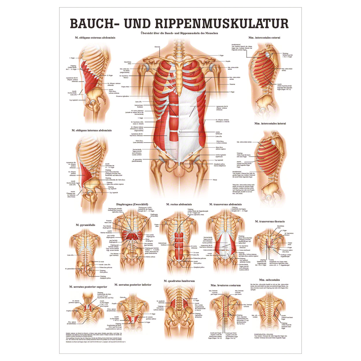
Poster Abdominal And Intercostal Muscles L X W 70x50 Cm Buy Online Sport Tec from www.sport-tec.com You'll need a bench and one dumbbell to do this exercise. Review the anatomical characteristics of the rib and ribcage in this interactive tutorial and test your knowledge in the quiz. It is formed by the vertebral column, ribs, and sternum and encloses the heart and lungs. Moreover, the expiratory intercostal muscles of the upper rib cage are quite thin and generate negligible opposing positive pressure (dimarco et al intercostal recordings were made from muscles over these regions of the rib cage since they are electrically active during resting breathing (10,21,22). Study flashcards on chapter 10 muscle diagrams at cram.com. Start studying rib cage muscles. As you inhale, the muscles in between the ribs lift the rib cage up, allowing the lungs to expand. Anatomical illustrations and diagrams of the spine (cervical, dorsal and lumbar) and back:
These muscles may be located anteriorly, posteriorly, and/or laterally.
The primary responsibilities of the ribcage involve protecting the thoracic visceral organs, enclosing the thoracic visceral organs, and is included in the general mechanics of the process of breathing. Start studying rib cage muscles. The ribs are curved, flat bones which form the majority of the thoracic cage. Unlike other cuff muscles, the rotatory component of the supraspinatus has a large enough moment arm (ma) that it is capable by itself producing a identify rib cage muscles. Anatomical illustrations and diagrams of the spine (cervical, dorsal and lumbar) and back: Muscles that move the rib cage attach to the rib cage. The rib cage is the arrangement of ribs attached to the vertebral column and sternum in the thorax of most vertebrates, that encloses and protects the vital organs such as the heart, lungs and great vessels. The following general rules regarding actions can be. They are attached to the femur (thighbone), tibia (shinbone), and fibula (calf bone) by fibrous tissues called ligaments. During normal breathing, the major inspiratory muscles produce rib cage expansion and a downward movement of the diaphragm. Perform dumbbell pullovers to work the muscles along your rib cage. There are twelve (12) pairs of ribs and all articulate posteriorly with the thoracic vertebrae. Study flashcards on chapter 10 muscle diagrams at cram.com.
Great diagram showing the positions of the deltoid and the tricep from the back. All muscles that are attached to the human rib cage have the inherent potential to cause a breathing action. Quickly memorize the terms, phrases and much more. • raise rib cage for inhaling & depresses rib cage for exhaling. Learn vocabulary, terms and more with flashcards, games and other study tools.
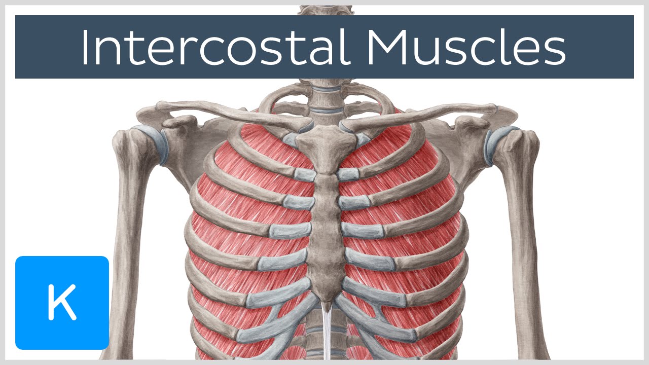
Intercostal Muscles Function Area Course Human Anatomy Kenhub Youtube from i.ytimg.com This is an online quiz called rib cage muscle diagram. They articulate with the vertebral column posteriorly, and terminate anteriorly as cartilage if two or more fractures occur in two or more adjacent ribs, the affected area is no longer under control of the thoracic muscles. Please click on the diagram(s) to view larger version. There is a printable worksheet available for download here so you can take the quiz with pen and paper. As you inhale, the muscles in between the ribs lift the rib cage up, allowing the lungs to expand. Muscles that move the rib cage attach to the rib cage. Unlike other cuff muscles, the rotatory component of the supraspinatus has a large enough moment arm (ma) that it is capable by itself producing a identify rib cage muscles. Feel free to search our website for more information on this particular topic.
When you exhale, the rib cage moves down again, squeezing the air.
These muscles may be located anteriorly, posteriorly, and/or laterally. Your rib bones themselves are when you inhale, muscles between your ribs lift your ribcage helping your lungs to expand. Diagram of human body, liver rib cage, rib cage diagram labeled, rib cage diagram numbered, rib cage diaphragm, rib cage heart, rib cage organs anatomy, rib cage pain, stomach. Your ribs form a protective cage that encloses many of your delicate internal organs, such as your heart and lungs. Tendons attach the muscles to each other. The two muscles which comprise the intermediate muscle group are the serratus posterior inferior, and the serratus posterior superior. 05.11.2019 · 16 photos of the rib cage diagram with organs diagram of human body, liver rib cage, rib cage diagram labeled, rib cage diagram numbered, rib cage diaphragm, rib cage heart. Introduction to the structure of the ribcage and ribs: It encloses and protects the heart and lungs. The rib cage has three important functions: Osteology, myology of the spine. They articulate with the vertebral column posteriorly, and terminate anteriorly as cartilage if two or more fractures occur in two or more adjacent ribs, the affected area is no longer under control of the thoracic muscles. Rib cage diagram this summary post is displaying rib cage diagram.
Muscles that move the rib cage attach to the rib cage. The other attachment of these muscles is usually considered to be either superior or inferior to the rib attachment. Learn vocabulary, terms and more with flashcards, games and other study tools. They are attached to the femur (thighbone), tibia (shinbone), and fibula (calf bone) by fibrous tissues called ligaments. The thoracic cage makes up the skeleton for the thoracic wall, and provides the attachments needed for the muscles of the neck, thorax.
Intercostal Muscles Diagram Quizlet from o.quizlet.com The rib cage is an arrangement of bones in the thorax of all vertebrates except the lamprey. The primary responsibilities of the ribcage involve protecting the thoracic visceral organs, enclosing the thoracic visceral organs, and is included in the general mechanics of the process of breathing. Muscles that helpful in expanding the thoracic cavity are called the inspiratory muscles because they help in inhalation, while those that compress the thoracic cavity are called expiratory. Check out our muscle anatomy reference charts to learn faster! Introduction to the structure of the ribcage and ribs: Muscles that move the rib cage attach to the rib cage. Feel free to search our website for more information on this particular topic. Learn vocabulary, terms and more with flashcards, games and other study tools.
The function of the rib cage is to filter the blood it receives, processing the blood.
• raise rib cage for inhaling & depresses rib cage for exhaling. As you inhale, the muscles in between the ribs lift the rib cage up, allowing the lungs to expand. Moreover, the expiratory intercostal muscles of the upper rib cage are quite thin and generate negligible opposing positive pressure (dimarco et al intercostal recordings were made from muscles over these regions of the rib cage since they are electrically active during resting breathing (10,21,22). Diagram of human body, liver rib cage, rib cage diagram labeled, rib cage diagram numbered, rib cage diaphragm, rib cage heart, rib cage organs anatomy, rib cage pain, stomach. When you exhale, your ribcage moves down, squeezing. Learn vocabulary, terms and more with flashcards, games and other study tools. Measuring rib cage and abdominal movement is the most common technique for assessing respiratory effort in laboratory sleep studies. Anatomical illustrations and diagrams of the spine (cervical, dorsal and lumbar) and back: Tendons attach the muscles to each other. Best viewed on 1280 x 768 px resolution in any modern browser. In humans, the rib cage, also known as the thoracic cage. The thoracic cage is part of the axial skeleton (also known as the rib cage), and consists of 24 ribs, the sternum, costal cartilage, and the 12 thoracic vertebrae. The other attachment of these muscles is usually considered to be either superior or inferior to the rib attachment.
Introduction to the structure of the ribcage and ribs: rib cage muscles. The primary responsibilities of the ribcage involve protecting the thoracic visceral organs, enclosing the thoracic visceral organs, and is included in the general mechanics of the process of breathing.


0 Komentar